Board Review: Down the watering hole
A 62 year old man presents to the ED with a chief complaint of acute onset severe flank pain radiating to the groin for the past hour. He has a history of HTN and tobacco use. Vital signs are T 37C HR 115 BP 110/80 RR 20 SpO2 99%. On exam, he appears uncomfortable with CVA tenderness on the left. Of the choices below, which imaging modality should you pursue first?
- CT with contrast
- CT without contrast
- POCUS
- XR
Flank pain and CVA tenderness is concerning for nephrolithiasis and/or pyelonephritis, and ultimately this patient requires CT imaging. However, a ruptured AAA can also present similarly to renal colic. Therefore, you should first perform a quick POCUS at the bedside to examine the aorta for this patient with significant risk factors and abnormal vital signs
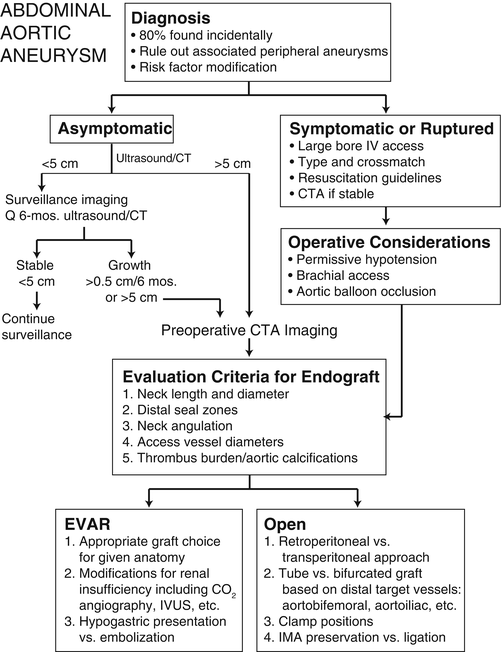
Aulivola B., Malinowski M. (2015) Abdominal Aortic Aneurysm. In: Saclarides T., Myers J., Millikan K. (eds) Common Surgical Diseases. Springer, New York, NY
See this prior EM Daily post about US findings for hydronephrosis, another possibility for this case: https://emdaily.cooperhealth.org/content/whats-diagnosis-dr-michael-tom-3