Critical Cases: smoke inhalation!
Patient:
30 yo F with PMH generalized anxiety disorder, presents to the ED after being in house fire. Unknown amount of time spent in burning house.
On presentation, she reports feeling like her throat is “smoky”. Otherwise, no symptoms, denies pain, sore throat, difficulty speaking, difficulty swallowing, shortness of breath and lightheadedness.
PE:
Vital signs: T 98.8, HR 120, BP, 152/70, RR 24, O2 sat 98% on 2L NC
Patient is awake, alert, speaking clearly, managing oral secretions
She has soot on her nose, cheeks, chin. No evidence of burns on her skin.
She has soot inside her nares and singed nasal hairs bilaterally.
Tachycardic with normal heart sounds.
Lungs are clear to auscultation.
Physical exam otherwise unremarkable.
What is the next step?
When seeing a patient who was in a fire, always consider not only burns on the skin, but also potential injury to the airway which may not be immediately visible on physical exam.
Smoke Inhalation Injury in the ED:
Symptoms of inhalation injury include: shortness of breath, sore throat, discomfort or swelling in mouth or throat
Signs of inhalation injury include: soot on the face or in the nares, singed nasal hairs, full thickness burns on the face, burns inside the mouth, supraglottic edema, neck swelling, tachypnea, hypoxia, respiratory distress.
Consider
-Airway management
-Possible exposure to toxic inhalants
Consider endotracheal intubation early! These patients have potential to progress to critical airway edema quickly as a result of inhaled particulate matter and direct thermal injury, which makes endotracheal intubation very difficult and potentially dangerous.
This patient presenting after being in a house fire, with no evidence of superficial burns, but with signs and symptoms concerning for possible inhalation injury.
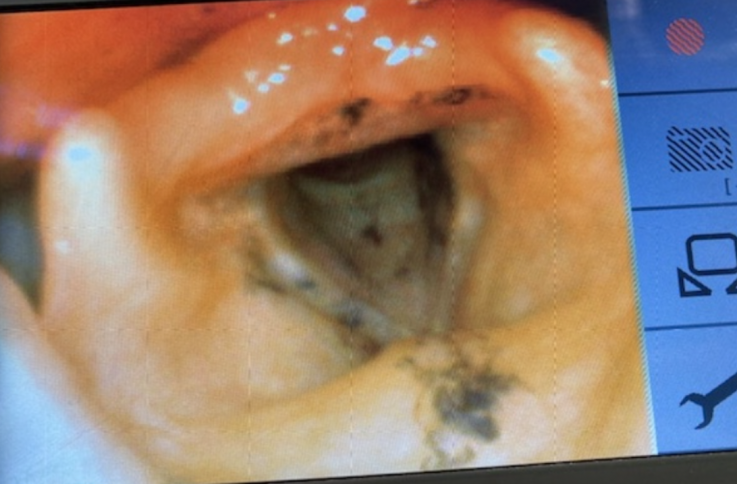
The patient underwent nasopharyngeal laryngoscopy in the ED, which showed no edema, but soot present on the epiglottis and vocal cords (see photo below). The decision was made to intubate the patient for airway protection in the setting on inhalation injury and possible progression to airway edema.
Consider Exposure to Toxic Inhalants
Carbon Monoxide
-CO binds to hemoglobin with affinity 200x that of oxygen, forming carboxyhemoglobin
-Carboxyhemoglobin does not participate in oxygen delivery to tissues, creates functional anemia and hypoxia; also impairs oxygen release to tissues from unaffected hemoglobin
-Pulse oximetry will be normal regardless of COHb level.
-Send carboxyhemoglobin level. Symptoms can be subtle, have low threshold to send test.
-If CO toxicity suspected or confirmed, apply oxygen in highest concentration possible and consider hyperbaric treatment depending on presentation (altered mental status, syncope, seizure, coma, high COHb levels, MI)
Cyanide
-Formed by combustion of nitrogen-containing polymers (wool, silk, plastics, which may be found in a home).
-Binds mitochondrial oxidative phosphorylation, inhibiting ATP production through aerobic cellular respiration, leading to profound tissue hypoxia and lactate production.
-Symptoms include dyspnea, restlessness, anxiety, palpitations, and headache. In significant inhalation exposure, can cause LOC, seizures, dysrhythmias, CV collapse.
-Send serum lactate level!! Levels over 10mmol/L are highly sensitive and specific for cyanide toxicity.
-If cyanide toxicity is suspected, treat with hydroxycobalamin or sodium nitrite + sodium thiosulfate.
References
Tintinalli’s Emergency Medicine: A Comprehensive Study Guide, 8th Edition. Ch 204, 216, 222. Judith E. Tintinalli.