Critical Cases - T Trach respiratory distress emergency!
Tue, 12/10/2019 - 5:54pm
Editor:
HPI:
- Pt had been complaining about abdominal pain earlier in the day
- Visiting nurse found pt in respiratory distress and covered in vomit
- On arrival to the ED, pt unable to provide much history due to tachypnea, nods "yes" as to whether his abdomen hurts
Pmhx:
- TBI, tracheal stenosis, tracheal malacia, subglottic stenosis, tracheostomy tube, ventral hernias, CVA, CAD
Exam
VS:
T: 97.5 (oral) HR: 145 (sinus) BP 176/92 RR 40 SpO2 92% on NRB
- Pt markedly tachypneic with abdominal retractions
- Oropharynx clear
- +tracheostomy tube in place, without an obvious balloon inflation port
- abd is distended, +diffusely tender
- +b/l LE edema
DDX
- Respiratory distress potentially d/t: vomiting with aspiration event, displaced trach, pneumonia, mucous plugging, atelectasis, pneumothorax, pulmonary embolism
- Abdominal pain: possible SBO leading to vomiting, possible incarcerated hernia, mesenteric ischemia
Initial resuscitation
- In general with tracheostomy emergences: 1) Aggressive in-line suctioning for possible mucous plugging, esp if poor air entry 2) apply high flow O2 to both the face and the trach 3) confirm trach placement with auscultation, capnometry, and/or direct visualization of the trachea/carina with a flexible endocscope advanced through tracheosotmy tube
- Airway equipment to bedside including video laryngoscope, intubating bronchoscope
- ENT called to assess trach
Initial studies

CXR interp: Slightly increased alveolar opacity at R base, consistent with possible aspiration
VBG:

VBG interpretation: Primary metabolic and primary respiratory acidosis
Management and Clinical Course
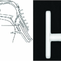
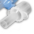
- Wife and ENT confirmed pt's trach to be a "T-trach" with a lumen that split after stoma entry, with one arm directed superior towards pts oropharynx and the other arm directed inferior to the trachea
- Pt's degree of respiratory distress precluded transport to CT for abdominal CT
- Decision made to attempt mechanical ventilation through T-Tube by attaching ET Tube adapter, however the majority of the tidal volume escaped through the patient's mouth

-
Decision made to replace T-tube with cuffed ET tube to allow for effective mechanical ventilation
- Pt was sedated without paralysis with ketamine
- Attempt made to pass ETT orotracheal failed due to subglottic stenosis
- T-Tube was pulled and quickly replaced with a cuffed 6.5 ET tube
- Tube placement confirmed with ET CO2 and a flexible intubating scope was also passed which visualized carina
Outcome
- Shortly after intubation, pt HR dropped and he experienced PEA cardiac arrest
- Was resuscitated and achieved ROSC
- Ultimately made comfort care and expired within 2 hours
- Likely cause of cardiac arrest was acidosis given concomitant presence of metabolic and respiratory acidosis, process of intubation likely caused worsening acidosis
Teaching Points
- Recognize the T-tube in tracheostomy patients, often placed to allow pts with both subglottic and tracheal stenosis to speak
- This tube is not cuffed and cannot be used for effective mechanical ventilation, the instilled breath will preferentially head towards the area of least resistance: the superior arm of the T-tube and out the mouth
- When replacing a tracheostomy tube, have all necessary equipment laid out and on hand
- Use video laryngoscopy and a flexible intubating scope to facilitate intubation through glottis or stoma and to confirm placement of tube along with CO2 capnometry