What's the Diagnosis? By Dr. Erica Schramm
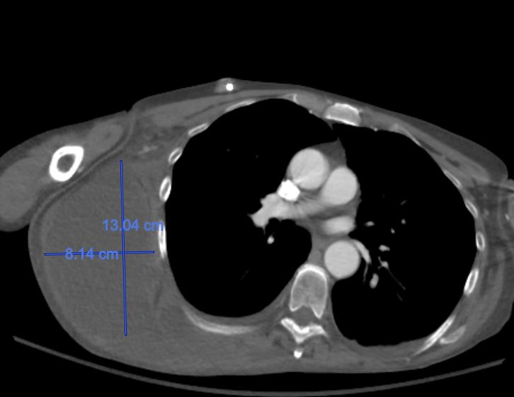
A 63 yo F w/ a history of colon cancer, prior PE (on lovenox) presents to the ED for a "mass" on her back x 3 days. The area has increased in size and become more painful. She denies trauma. She is well appearing and hemodynamically stable. Labs are significant for a Hgb of 4.3 and an INR of 2.3. A CT scan of the chest/abdomen/pelvis is shown below. What's the diagnosis? (scroll down for answer)
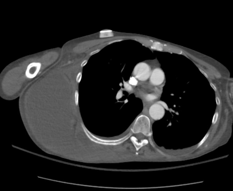
Answer: Spontaneous hematoma of the right chest/abdominal wall
- Rare occurrence, rarely reported in medical literature outside of case reports
- Therapeutic anticoagulation is major risk factor
- Intervention varies and is directed by hemodynamic stability of the patient (medical management includes blood transfusion, serial Hgb/Hct; surgical intervention involves hematoma evacuation/control of bleeding)
- The patient above was managed conservatively with PRBC/FFP transfusion, and repeat Hgb/INR improved
- Etiology of bleeding was likely related to patient's anticoagulation
References:
1) Bevan P, Menon A, and Bunton R. Spontaneous Chest Wall Hematoma with Dual Antiplatelet Therapy. Canadian Journal of Cardiology; Volume 30, Issue 2, 2014
2) Salemis N. Rivaroxaban-induced Chest Wall Spontaneous Expanding Hematoma. Drug Discoveries & Therapeutics; Volume 11, Issue 1, 2017