What's the Diagnosis? By Dr. Katie Selman
A 63 yo F is brought in by EMS after being found down. She has multiple ecchymoses on her chest and b/l flanks. GCS is 6. After intubation, she is taken for CT head/cervical spine and a CT chest/abdomen/pelvis with contrast. Upon return from CT, X-rays are done (shown below) to further evaluate bruising and a laceration to her L elbow. What's the diagnosis? (scroll down for answer)
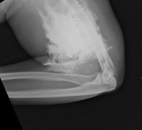
Answer: Contrast extravasation
- Predisposing factors for contrast extravasation
- Small IV gauge (22G or less)
- More distal access (hand)
- Rapid injection of contrast
- Incidence: up to 1% of patient receiving IV contrast through peripheral IV
- Most common symptoms: local pain, swelling
- Complications occur in < 1 % (more common with large volume and in patients with atherosclerosis, venous insufficiency, or impaired lymphatic drainage)
- Compartment syndrome
- Tissue necrosis
- Close monitoring required following extravasation
- Compartment checks, vascular checks, and monitoring of overlying skin
- Surgery consult for any signs of compartment syndrome or tissue injury
- Elevate limb, warm compresses may be used
- Patients rarely require more than conservative supportive treatment
References:
Sbitany, H., Koltz, P. F., Mays, C., Girotto, J. A., & Langstein, H. N. (2010). CT contrast extravasation in the upper extremity: Strategies for management. International Journal of Surgery, 8(5), 384-386. doi:10.1016/j.ijsu.2010.06.002
Sonis, J. D., et al (2018). Implications of iodinated contrast media extravasation in the emergency department. The American Journal of Emergency Medicine, 36(2), 294-296. doi:http://dx.doi.org/10.1016/j.ajem.2017.11.012