What's the Diagnosis? By Dr. Rebecca Fieles
Wed, 09/02/2020 - 3:00pm
Editor:
A 44 yo M presents to the ED w/ L foot and ankle pain. He was running and stepped into a hole, stating he heard a "crack". He has been unable to bear weight since the injury. On exam, he has swelling and marked bony tenderness of both the lateral and medial malleoli and heel. Xrays of the L foot were obtained. What's the diagnosis? (scroll down for answer)
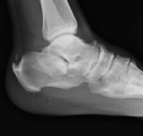
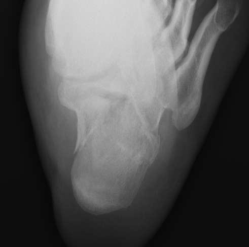
Answer: Comminuted fracture of calcaneus
- Etiology
- Most commonly due to high axial load injuries such as fall from height or MVC
- ** most common tarsal fracture
- Presentation
- Diffuse pain, swelling and ecchymosis after trauma
- Often unable to bear weight
- Deformity of heel or plantar arch on exam
- Mondor's sign- ecchymosis/hematoma that tracks along sole of foot (pathognomonic for calcaneal fracture)
- Diagnosis
- Plain radiographs of ankle/foot
- Harris view: calcaneus in axial view
- Non-contrast CT of the foot/ankle is the gold standard and assists with surgical planning
- Sander's Classification (based on CT)
- Type I: all intra-articular fractures that have < 2mm displacement, regardless of # of fracture lines/fragments
- Type II: two bony fragments involving posterior facet
- Type III: three bony fragments including depressed middle fragment
- Type IV: four comminuted bony fragments
- ED management
- assess NV status
- analgesia, ice, elevation
- splinting, often with bulky Jones dressing
- ortho consult
- Most intra-articular fractures require surgical repair
- Most extra-articular fractures can be treated conservatively with 10-12 weeks of casting; pt discharged from ED non-weightbearing
References:
Jiménez-Almonte JH, King JD, Luo TD, Aneja A, Moghadamian E. Classifications in Brief: Sanders Classification of Intraarticular Fractures of the Calcaneus. Clin. Orthop. Relat. Res. 2019 Feb;477(2):467-471